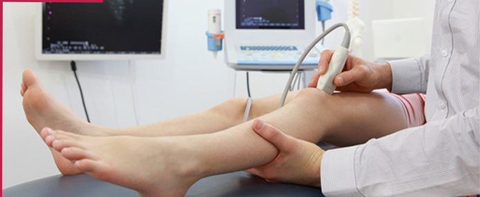
An ultrasound-guided needle biopsy is a medical test used to learn more about a lesion or mass. The biopsy is done by using an ultrasound to find the lesion or mass. This is one type of "image-guided" biopsy, which combines the use of ultrasound with either a Fine Needle Aspiration or Core Needle Biopsy. This test is most often used for lymph node, breast, and liver biopsies. Fine-needle aspiration biopsy (FNAB) was utilized for many years to investigate the breast tissue in the attempt to avoid surgical biopsy (gold standard). With the arrival of core biopsy, a better specimen quality could be obtained and it became possible to differentiate carcinomas in situ from invasive carcinomas.
In cases of breast lesions, core biopsy (CB) is preferably performed, utilizing an imaging method as guidance - for example: ultrasonography (US) or stereotactic biopsy -, but it is still performed, with lower sensitivity, only by means of palpation. The present study is aimed at detailing the main steps of US-guided CB of breast, including indications, advantages, limitations, follow-up and description of the technique, besides presenting a checklist including the critical steps required for an appropriate practice of such procedure.

An ultrasound-guided breast biopsy uses sound waves to help locate a lump or abnormality and remove a tissue sample for examination under a microscope. It is less invasive than surgical biopsy, leaves little to no scarring and does not involve exposure to ionizing radiation.

Success using the pigtail catheter demands adherence to proper patient selection and attention to details at the time of insertion. A lateral decubitus chest radiograph is a rapid and simple way of assuring that an effusion is free-flowing and, therefore, likely to respond to pigtail drainage. Small-bore chest tubes – also referred to as pigtail catheters – are being used to relieve both spontaneous and in some cases, traumatic pneumothorax. Thesepigtails are placed with a Seldinger catheter-over-wire technique very similar to the central venous catheter insertion.
Other tests that you have probably had, such as an ultrasound or CT scan, will have shown that you have a collection and that it is suitable for draining through a small tube, rather than by an open operation. Once this fluid has been removed, you should feel more comfortable. Sometimes a sample of the fluid that has been removed will be sent to the laboratory for testing, which means we can diagnose and treat your symptoms more effectively.

Ultrasound- and MRI-guided prostate biopsies are performed to collect tissue samples from the prostate gland for examination by a pathologist to determine whether or not the tissue is cancerous. Biopsies are most commonly performed under ultrasound guidance. During the procedure, a special biopsy needle is inserted into the prostate gland through the wall of the rectum to remove several small samples of tissue for pathologic analysis. This method is known as transrectal ultrasound (TRUS) guided biopsy.
The prostate may also be accessed through the perineum (the area of skin between the base of the penis and the rectum). This method is known as the transperineal approach and may be used for one of several reasons:
Transrectal ultrasound guided (TRUS) biopsy. Doctors use this test to diagnose prostate cancer. They take samples of tissue from the prostate gland to look for cancer cells. You might have an MRI scan before your transrectal ultrasoundguided biopsyA prostate biopsy is a procedure to remove samples of suspicious tissue from the prostate. The prostate is a small, walnut-shaped gland in men that produces fluid that nourishes and transports sperm. During a prostate biopsy a needle is used to collect a number of tissue samples from your prostate gland. The procedure is performed by a doctor who specializes in the urinary system and men's sex organs (urologist). Your urologist may recommend a prostate biopsy if results from initial tests, such as a prostate-specific antigen (PSA) blood test or digital rectal exam, suggest you may have prostate cancer. Tissue samples from the prostate biopsy are examined under a microscope for cell abnormalities that are a sign of prostate cancer. If cancer is present, it is evaluated to determine how quickly it's likely to progress and to determine your best treatment options.
Prostate biopsy samples can be collected in different ways. Your prostate biopsy may involve:
Success using the pigtail catheter demands adherence to proper patient selection and attention to details at the time of insertion. A lateral decubitus chest radiograph is a rapid and simple way of assuring that an effusion is free-flowing and, therefore, likely to respond to pigtail drainage. Small-bore chest tubes – also referred to as pigtail catheters – are being used to relieve both spontaneous and in some cases, traumatic pneumothorax. Thesepigtails are placed with a Seldinger catheter-over-wire technique very similar to the central venous catheter insertion.
Other tests that you have probably had, such as an ultrasound or CT scan, will have shown that you have a collection and that it is suitable for draining through a small tube, rather than by an open operation. Once this fluid has been removed, you should feel more comfortable. Sometimes a sample of the fluid that has been removed will be sent to the laboratory for testing, which means we can diagnose and treat your symptoms more effectively.

Percutaneous transhepatic biliary drainage (PTBD) is a common and effective procedure for the palliation of cholestasis in both malignant and benign biliary obstruction, especially after the application of the B-mode ultrasonography guidance . Success rates for PTBD have been reported at 90% or more, and complication rates have been reported at 3% or less .
However, when applied to patients with a special condition (for example, a nondilated bile duct, patient status post left hepatic lobe resection, patient status post liver transplantation), PTBD has the potential for technical difficulties. In addition, for new practitioners of this procedure, understanding the bile duct anatomy (especially the right hepatic bile duct) can be challenging. A procedure to drain bile to relieve pressure in the bile ducts caused by a blockage. An x-ray of the liver and bile ducts locates the blockage of bile flow. Images made by ultrasound guide placement of a stent (tube), which remains in the liver. Bile drains through the stent into the small intestine or into a collection bag outside the body. This procedure may relieve jaundice before surgery. Also called percutaneous transhepatic cholangiodrainage and PTCD.

A percutaneous nephrostomy is the placement of a small, flexible rubber tube(catheter) through your skin into your kidney to drain your urine. Percutaneousnephrostolithotomy (or nephrolithotomy) is the passing of a special medical instrument through your skin into your kidney. This is done to remove kidney stones. Most stones pass out of the body on their own through urine. When they do not, your health care provider may recommend these procedures. During the procedure, you lie on your stomach on a table. You are given a shot of lidocaine. This is the same medicine your dentist uses to numb your mouth. The provider may give you medicines to help you relax and reduce pain. If you have nephrostomy only:
Procedure: A retrograde pyelogram is done to locate the stone in the kidney. With a small 1 centimeter incision in the loin, the percutaneous nephrolithotomy (PCN) needle is passed into the pelvis of the kidney. The position of the needle is confirmed by fluoroscopy. A guide wire is passed through the needle into the pelvis. The needle is then withdrawn with the guide wire still inside the pelvis. Over the guide wire the dilators are passed and a working sheath is introduced. A nephroscope is then passed inside and small stones taken out. In case the stone is big it may first have to be crushed using ultrasound probes and then the stone fragments removed. The most difficult portion of the procedure is creating the tract between the kidney and the flank skin. Most of the time this is achieved by advancing a needle from the flank skin into the kidney, known as the 'antegrade' technique. A 'retrograde' technique has recently been updated wherein a thin wire is passed from inside the kidney to outside the flank with the aid of a flexible ureteroscope. This technique may reduce radiation exposure for patient and surgeon